Demystifying Hypoechoic Tumors: Causes and Treatments
Welcome to our blog post on hypoechoic nodules! If you’re here, chances are you’ve heard this term before and have some questions about what it means and how it relates to your health. Well, you’ve come to the right place! In this article, we’ll dive deep into the world of hypoechoic nodules – their definition, characteristics, potential causes, symptoms, diagnosis methods, treatment options, lifestyle modifications, and more. Whether you’re a medical professional or simply someone looking for information on this topic, we aim to provide you with valuable insights that will help you better understand hypoechoic nodules and make informed decisions about your health. So let’s get started on this enlightening journey together!
What is a Hypoechoic Nodule?
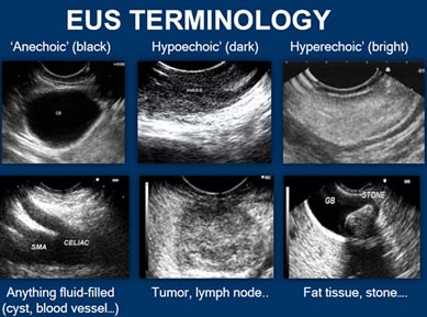
A hypoechoic nodule refers to an abnormality or growth that appears darker on ultrasound imaging compared to the surrounding tissues. These nodules can occur in various parts of the body, but are commonly associated with thyroid conditions. When an ultrasound is performed, sound waves are used to create images of the internal structures. Different types of ultrasound images help visualize different characteristics of tissues and organs. In the case of hypoechoic nodules, they appear as dark or hypoechoic areas on the ultrasound image, indicating a difference in density or composition compared to normal tissue. This distinct appearance helps medical professionals identify and monitor these nodules for potential health concerns.
Understanding what hypoechoic nodules are and how they present on ultrasound imaging plays a crucial role in their diagnosis and management.
Definition and Characteristics
A hypoechoic nodule refers to an abnormal growth or mass in the body that appears darker or less echoic on ultrasound imaging. These nodules have distinct characteristics that help differentiate them from surrounding tissues. They typically have a solid composition, irregular borders, and can vary in size.
Hypoechoic nodules are often associated with changes in tissue density and can occur in various organs such as the thyroid gland, breast, liver, or kidney. These nodules may be indicative of underlying health issues and require further evaluation to determine their nature and potential risk factors. Ultrasound imaging plays a crucial role in identifying these nodules and providing valuable information for diagnosis and treatment planning.
How Ultrasound Works
Ultrasound is a widely used diagnostic tool that plays a crucial role in identifying hypoechoic nodules. But have you ever wondered how ultrasound actually works? Well, it’s quite fascinating!
During an ultrasound exam, a transducer emits high-frequency sound waves into the body. These waves then bounce off different tissues and organs, creating echoes that are captured by the transducer. The echoes are then processed by a computer to create real-time images on a monitor. This allows healthcare professionals to visualize the size, shape, and composition of any abnormal structures or nodules present in the thyroid gland. In essence, ultrasound provides valuable insight into what’s going on inside your body without using radiation or invasive procedures.
So while it may seem like magic when you see those detailed images of your thyroid on the screen during an ultrasound examination, it’s actually science at work! Ultrasound technology has revolutionized medical imaging by providing non-invasive and safe ways to evaluate various conditions like hypoechoic nodules in order to make accurate diagnoses and determine appropriate treatment plans. It truly is remarkable how far we’ve come in our ability to see beneath the surface of our skin!
Types of Ultrasound Images
Ultrasound imaging plays a crucial role in diagnosing and monitoring hypoechoic nodules. There are different types of ultrasound images that provide valuable insights into the characteristics of these nodules.
One type is the B-mode ultrasound, which uses grayscale to create a detailed image of the nodule’s shape, size, and boundaries. This helps doctors determine if the nodule is solid or cystic. Another type is the Doppler ultrasound, which evaluates blood flow within and around the nodule. By analyzing vascularity patterns, it can help identify potential malignancies.
These various types of ultrasound images enable healthcare professionals to assess hypoechoic nodules comprehensively and make informed decisions regarding further diagnostic procedures or treatment options. Understanding these images allows for accurate evaluation and management of patients with suspected or diagnosed nodules.
Are Hypoechoic Nodules Cancerous?
Understanding the Risk of Cancer
When it comes to hypoechoic nodules, one question that often arises is whether or not they are cancerous. While hypoechoic nodules can be a cause for concern, it’s important to understand the risk of cancer associated with them.
Differentiating Benign vs Malignant Nodules
Not all hypoechoic nodules are cancerous. In fact, most hypoechoic nodules are benign, meaning they are non-cancerous. However, there is still a possibility that a hypoechoic nodule could be malignant and indicate thyroid cancer. It’s essential to differentiate between benign and malignant nodules through further testing and evaluation by healthcare professionals.
Understanding the Risk of Cancer
When it comes to hypoechoic nodules, one of the primary concerns is determining whether they are cancerous or not. While not all hypoechoic nodules are malignant, it’s crucial to understand the risk involved. The presence of certain factors can increase the likelihood of a nodule being cancerous.
Factors such as age, family history of thyroid cancer, and exposure to radiation may raise concern. Additionally, characteristics observed during an ultrasound examination can provide valuable insight into the potential malignancy of a nodule. It’s important to consult with healthcare professionals who specialize in thyroid conditions for accurate assessment and appropriate management strategies. Stay informed about your individual risk factors and work closely with your medical team to ensure proper monitoring and timely intervention if needed.
Remember that understanding the risk associated with hypoechoic nodules is essential for making informed decisions about your health. By staying proactive and seeking professional guidance when necessary, you can take steps towards managing these concerns effectively.
Differentiating Benign vs Malignant Nodules
Understanding the difference between benign and malignant nodules is crucial in determining the appropriate course of action. Benign nodules are non-cancerous growths that often do not require immediate treatment. On the other hand, malignant nodules are cancerous and may require prompt intervention. Differentiating between the two can be challenging as they may share similar characteristics on ultrasound images. However, additional diagnostic methods such as blood testing and fine-needle biopsy can provide more accurate results to determine whether a nodule is benign or malignant.
In some cases, benign nodules have distinct features that indicate their non-cancerous nature. These features include regular borders, uniform texture, absence of calcification, and stable size over time. Malignant nodules, on the other hand, may exhibit irregular borders with indistinct edges, heterogeneous texture with areas of hypoechoic appearance (lower echogenicity than surrounding tissue), presence of calcifications or microcalcifications (tiny calcium deposits), and an increase in size over time. By carefully analyzing these characteristics along with additional diagnostic approaches, healthcare professionals can make an informed decision regarding further treatment options for patients with hypoechoic nodules.
Causes of Hypoechoic Nodules
Hypoechoic nodules can be caused by various factors, including thyroid conditions and other medical issues. Thyroid conditions such as Hashimoto’s disease or Graves’ disease can lead to the development of these nodules. These conditions cause inflammation or abnormal growth in the thyroid gland, resulting in hypoechoic areas on ultrasound images.
In addition to thyroid conditions, there are other medical factors that may contribute to the formation of hypoechoic nodules. One example is cysts, which are fluid-filled sacs that can develop in different parts of the body. Another possible cause is a goiter, which is an enlarged thyroid gland. It’s important to note that certain risk factors like age, gender, family history, and exposure to radiation may also increase the chances of developing hypoechoic nodules.
Remember: always consult with a healthcare professional for accurate diagnosis and treatment options based on your specific situation!
Thyroid Conditions
Thyroid conditions can play a significant role in the development of hypoechoic nodules. The thyroid gland, located at the base of the neck, produces hormones that help regulate our metabolism and energy levels. When this delicate balance is disrupted, it can lead to various thyroid conditions.
One common condition is called Hashimoto’s disease, an autoimmune disorder where the immune system attacks the thyroid gland. Another condition is Graves’ disease, which causes overproduction of thyroid hormones. These conditions can increase the risk of developing hypoechoic nodules and should be monitored closely by healthcare professionals.
Other Medical Conditions
Aside from thyroid conditions, there are other medical conditions that can contribute to the development of hypoechoic nodules. These include autoimmune disorders such as Hashimoto’s thyroiditis or Graves’ disease. Inflammation of the thyroid gland, known as thyroiditis, can also lead to the formation of hypoechoic nodules.
Additionally, certain infectious diseases like tuberculosis and fungal infections may be associated with the appearance of hypoechoic nodules in the thyroid gland. It’s important to note that these conditions should be diagnosed and treated properly by a healthcare professional to prevent further complications.
Potential Risk Factors
Certain factors may increase the risk of developing hypoechoic nodules. While these risk factors do not guarantee the presence of a nodule, they should be taken into consideration when assessing one’s overall health.
Thyroid conditions, such as Hashimoto’s thyroiditis or Graves’ disease, have been associated with an increased likelihood of developing hypoechoic nodules. Additionally, other medical conditions like iodine deficiency or radiation exposure to the neck area can also contribute to their formation. It is important to note that each individual may respond differently to these risk factors, and further research is needed to fully understand their impact on nodule development.
Symptoms and Diagnosis
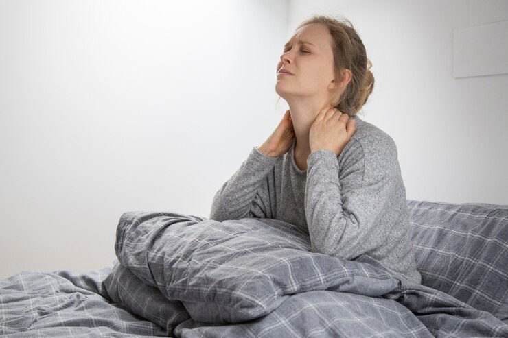
Recognizing Symptoms
When it comes to hypoechoic nodules, being aware of potential symptoms is crucial for early detection. Keep an eye out for any changes in your throat, such as a lump or swelling. Pay attention to any difficulty swallowing or breathing, as well as hoarseness or voice changes. Fatigue and unexplained weight loss may also be signs that something isn’t quite right.
Diagnostic Methods
If you experience any of these symptoms or have concerns about hypoechoic nodules, it’s important to seek medical advice promptly. Your healthcare provider may recommend various diagnostic methods to assess the nodules further. These can include blood testing to check hormone levels and fine-needle biopsy to collect tissue samples for analysis. By undergoing these tests, you can gain a better understanding of your condition and determine the appropriate course of action moving forward.
Recognizing Symptoms
When it comes to hypoechoic nodules, being aware of the symptoms is crucial. While some individuals may not experience any noticeable signs, others may have certain indicators that something isn’t quite right in their thyroid gland. Pay close attention to any changes in your body, such as a lump or swelling in your neck region. Additionally, keep an eye out for difficulty swallowing or breathing, hoarseness or voice changes, and unexplained weight loss or gain.
It’s important to remember that these symptoms can be caused by various other conditions as well and may not necessarily indicate the presence of hypoechoic nodules. However, if you do notice any of these signs persisting or worsening over time, it’s essential to consult with a healthcare professional for further evaluation and diagnosis. Early detection and prompt medical attention can make all the difference in managing thyroid health effectively. Stay vigilant!
Diagnostic Methods
When it comes to diagnosing hypoechoic nodules, recognizing the symptoms is crucial. While some individuals may not experience any noticeable signs, others might notice a lump or swelling in their neck area. Additionally, if you’re experiencing difficulty swallowing or breathing, it could be an indication of a more advanced nodule.
To confirm the presence of hypoechoic nodules and assess their characteristics, doctors rely on various diagnostic methods. One common approach is performing an ultrasound scan of the thyroid gland. This non-invasive imaging technique uses sound waves to create detailed images of the thyroid and identify any abnormalities present. Blood testing can also provide valuable information about hormone levels and potential underlying conditions that may contribute to nodule development. In cases where further evaluation is needed, a fine-needle biopsy may be performed to extract cells from the nodule for microscopic analysis. These diagnostic methods help healthcare professionals accurately diagnose and determine appropriate treatment plans for patients with hypoechoic nodules.
Blood Testing
Recognizing symptoms of hypoechoic nodules is crucial for early diagnosis and treatment. However, in order to confirm the presence of these nodules and determine their nature, blood testing may be necessary. Blood tests can provide valuable information about the functioning of the thyroid gland and help identify any abnormalities or imbalances in hormone levels. This can aid doctors in making an accurate diagnosis and developing an appropriate treatment plan.
During a blood test, a small sample of blood is taken from the patient’s arm and sent to a laboratory for analysis. The lab technicians will assess various markers such as thyroid-stimulating hormone (TSH), thyroxine (T4), triiodothyronine (T3), and antibodies that are associated with certain thyroid conditions. These results can indicate whether there are any underlying issues contributing to the development of hypoechoic nodules. Blood testing is a non-invasive procedure that offers valuable insights into your overall thyroid health, helping healthcare professionals make informed decisions regarding further diagnostic procedures or treatment options.
Fine-Needle Biopsy
Fine-Needle Biopsy, a commonly used diagnostic method for hypoechoic nodules, involves the extraction of tissue samples using a thin needle. This procedure is usually performed under ultrasound guidance to ensure accuracy. It is a minimally invasive technique that allows doctors to examine the nodule and determine if it is benign or malignant.
During the biopsy, a small amount of local anesthesia may be administered to minimize discomfort. The needle is then inserted into the nodule, and several samples are collected for laboratory analysis. Fine-needle biopsy provides valuable information about the composition and nature of the nodule, aiding in diagnosis and treatment planning without requiring surgery.
Treatment Options
When it comes to treating hypoechoic nodules, there are a few different options to consider. One approach is the “wait and watch” method, where doctors closely monitor the nodule over time to see if any changes occur. This can be appropriate for small nodules that are not causing any symptoms or health concerns.
In some cases, medication and supplements may be prescribed to help manage the underlying condition causing the nodule. These medications can help regulate hormone levels and reduce inflammation in the thyroid gland. In more severe cases or when nodules are deemed cancerous, surgical interventions such as thyroidectomy may be necessary to remove all or part of the thyroid gland. The specific treatment plan will depend on various factors including the size of the nodule, its characteristics, and individual patient factors
Wait and Watch Approach
For some individuals diagnosed with hypoechoic nodules, a “wait and watch” approach may be recommended by healthcare professionals. This approach involves monitoring the size and growth of the nodule over time without immediately intervening with treatment. It is often used for small nodules that are not causing any symptoms or posing an immediate risk.
By closely observing the nodule through regular follow-up appointments and imaging tests, doctors can assess whether any changes occur in its size or appearance. This allows them to make informed decisions regarding further interventions if necessary. The wait and watch approach can provide valuable insights into the behavior of hypoechoic nodules and help determine if additional steps need to be taken for proper management.
Medication and Supplements
Medication and supplements can play a role in managing hypoechoic nodules. Depending on the specific condition, your healthcare provider may prescribe medications to help regulate thyroid hormone levels or reduce inflammation. These medications can help alleviate symptoms and potentially shrink the size of the nodules over time.
In addition to medication, certain dietary supplements may also be recommended. Supplements like iodine, selenium, and vitamin D have been studied for their potential benefits in thyroid health. However, it’s important to consult with your doctor before starting any new supplement regimen as they can interact with other medications or have unintended side effects.
Surgical Interventions
When it comes to treating hypoechoic nodules, surgical interventions may be considered as an option. In some cases, surgery may be necessary if the nodule is causing significant symptoms or if there is a high suspicion of cancer.
During the surgical procedure, the nodule or part of the thyroid gland may be removed. This can help alleviate any discomfort and reduce the risk of potential complications. However, it’s important to remember that surgery is not always required and should only be pursued after careful consideration and consultation with a healthcare professional who will assess your individual case and make personalized recommendations for your situation.
Lifestyle Modifications for Hypoechoic Nodules
When it comes to managing hypoechoic nodules, making certain lifestyle modifications can play a crucial role in promoting overall well-being. Paying attention to your diet is important. Incorporating nutrient-rich foods like fruits, vegetables, whole grains, and lean proteins can provide the necessary vitamins and minerals for a healthy thyroid.
In addition to dietary changes, stress management techniques are essential. High levels of stress have been linked to various health issues including thyroid disorders. Engaging in activities like meditation, deep breathing exercises or yoga can help reduce stress levels and promote relaxation. Regular exercise is also beneficial as it not only helps maintain a healthy weight but also boosts energy levels and supports optimal thyroid function.
Remember that these lifestyle modifications should be tailored to individual needs and preferences. Consulting with a healthcare professional or registered dietitian can provide personalized guidance on implementing these changes effectively.
Dietary Changes
When it comes to managing hypoechoic nodules, making some simple dietary changes can play a significant role in supporting your overall thyroid health. One of the key factors is ensuring adequate iodine intake, as this mineral is essential for proper thyroid function. Including iodine-rich foods like seaweed, fish, and dairy products can help maintain optimal levels.
Additionally, incorporating selenium-rich foods such as Brazil nuts, eggs, and legumes into your diet may also be beneficial. Selenium plays a crucial role in the production of thyroid hormones and has antioxidant properties that protect the thyroid gland from damage.
Remember to consult with your healthcare provider or a registered dietitian before making any drastic dietary changes to ensure they align with your individual needs and medical condition. Making informed choices about what you eat can be an empowering step towards supporting your thyroid health.
Stress Management
When dealing with hypoechoic nodules, it’s important to address the impact of stress on your overall well-being. Stress can have a negative effect on your body and mind, exacerbating symptoms and potentially hindering recovery. Finding effective ways to manage stress is crucial.
One way to manage stress is through relaxation techniques such as deep breathing exercises or meditation. Taking time out of your day to practice these techniques can help calm the mind and reduce anxiety levels. Additionally, engaging in activities that bring you joy and provide an outlet for stress, such as hobbies or spending time in nature, can be beneficial for managing stress levels associated with hypoechoic nodules.
Exercise and Physical Activity
Regular exercise and physical activity are essential components of a healthy lifestyle, especially when dealing with hypoechoic nodules. Engaging in physical activities can help improve overall thyroid function and promote optimal health. Whether it’s going for a brisk walk, practicing yoga, or hitting the gym, finding an exercise routine that suits your preferences is key.
Exercise not only helps maintain a healthy weight but also boosts metabolism and improves blood circulation. It can also reduce stress levels and support mental well-being. Remember to start slowly if you’re new to exercising and gradually increase intensity over time. Always consult with your healthcare provider before starting any new exercise regimen to ensure it aligns with your specific needs and limitations. Stay active, stay healthy!
Important Considerations and Precautions
Follow-up monitoring is essential when it comes to hypoechoic nodules. Regular check-ups with your healthcare provider are crucial to keep track of any changes in the size or characteristics of the nodule. This will help determine whether further interventions or treatments are necessary.
It’s also important to be aware of potential complications that may arise from hypoechoic nodules. While most nodules are benign, there is always a small risk of malignancy. Therefore, staying vigilant and seeking medical attention if you experience any new symptoms or significant changes is vital for early detection and timely intervention if needed. Remember, taking care of your health should always be a priority!
Follow-up Monitoring
Regular monitoring is crucial when it comes to hypoechoic nodules. After an initial diagnosis, your doctor will likely recommend ongoing follow-up appointments to closely monitor any changes in the size or characteristics of the nodule.
During these check-ups, your doctor may perform repeat ultrasounds and other diagnostic tests to assess any potential growth or development. They will also evaluate if there are any new symptoms or concerning signs that need further investigation. By staying vigilant with regular follow-up monitoring, you can ensure early detection and timely intervention if necessary.
Remember, even though most hypoechoic nodules are benign, it’s important not to become complacent. Regular check-ups provide peace of mind and enable swift action should anything change in your thyroid health. Stay committed to your follow-up appointments and maintain open communication with your healthcare team for optimal management of hypoechoic nodules.
Potential Complications
It’s important to be aware of the potential complications that can arise from hypoechoic nodules. While most nodules are benign and don’t cause any significant problems, there is a small chance that they could develop into cancerous growths. This is why regular monitoring and follow-up appointments with your healthcare provider are crucial.
Additionally, if the nodule grows larger or puts pressure on surrounding structures, it can lead to symptoms such as difficulty swallowing or breathing. In rare cases, a large nodule may even cause compression of the windpipe or esophagus, requiring immediate medical attention. It’s essential to stay vigilant and report any new symptoms or changes in your condition to your doctor promptly.
Importance of Regular Check-ups
Regular check-ups are a crucial aspect of managing hypoechoic nodules. By staying proactive and monitoring any changes in the thyroid gland, you can catch potential issues early on and ensure timely intervention if necessary.
These routine check-ups allow healthcare professionals to assess the size, shape, and characteristics of the nodules through ultrasound imaging or other diagnostic methods. They can also conduct blood tests to measure hormone levels and evaluate overall thyroid function.
Regular check-ups serve as a valuable opportunity for doctors to track the progression of your condition over time. By comparing previous imaging results with current ones, they can identify any significant changes that may warrant further investigation or treatment.
Furthermore, regular check-ups enable healthcare providers to address any concerns or symptoms you may have promptly. This ongoing communication helps build a strong doctor-patient relationship based on trust and collaboration.
Remember that even if your hypoechoic nodule is initially deemed benign or low-risk, it’s essential not to neglect follow-up appointments. Nodules can change over time, potentially becoming larger or more suspicious-looking. Timely detection of these changes allows for appropriate interventions before complications arise.
In conclusion (without explicitly stating it), regular check-ups play a vital role in effectively managing hypoechoic nodules by ensuring close monitoring and timely intervention when needed. So make sure to prioritize these visits as part of your overall health maintenance plan!